Baker's Cyst: That Lump Behind Your Knee — What It Is and Why It Keeps Coming Back

Most lumps behind the knee are Baker's cysts fed by excess fluid from inside the joint.
A Baker's cyst — more formally a popliteal cyst — is a fluid-filled swelling that develops behind the knee. It feels like a tense, sometimes tender lump in the crease of the back of the knee. It may cause a pulling or tightness sensation when straightening the leg. Occasionally it is dramatic enough to restrict bending.
Patients find them by accident, feel them during exercise, or notice them when a family member spots the swelling. The first instinct — naturally — is to worry. The second instinct is to look up "lump behind knee" and spiral through cancer concerns until they see a doctor.
The reassurance, which should come first: the vast majority of popliteal lumps in adults are Baker's cysts, and Baker's cysts are benign. They are not cancer. They do not become cancer. The clinical concern is not the cyst itself — it is the underlying joint problem that is producing the excess fluid the cyst contains.
What a Baker's Cyst Actually Is
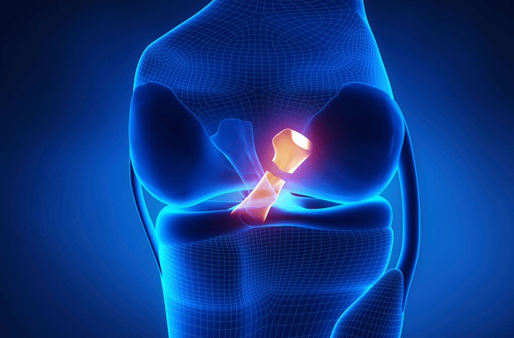
The knee joint is enclosed in a capsule lined by synovium — the membrane that produces synovial fluid (joint lubrication). At the back of the knee, the capsule has a natural weak point between the medial head of the gastrocnemius (the inner calf muscle) and the semimembranosus tendon. In some people, this creates a one-way valve: fluid from the joint can flow back into the bursa (a fluid-filled sac) in this space, but cannot easily return.
When anything causes increased production of synovial fluid — which is the body's response to inflammation inside the joint — this excess fluid can accumulate in the popliteal bursa, forming the characteristic cyst.
The cyst does not arise from nowhere. It is a symptom of something happening inside the knee joint. The most common causes:
Osteoarthritis of the knee: The inflamed arthritic joint produces excess synovial fluid. This is the most common cause of Baker's cyst in adults over 50.
Meniscal tears: A torn meniscus triggers inflammation that increases fluid production. Baker's cysts in younger adults are frequently associated with meniscal pathology.
Rheumatoid arthritis: Active RA in the knee produces significant synovitis and excess fluid; Baker's cysts are common in RA.
Non-specific synovitis: Inflammatory flares without a specific identified structural cause.
How to Distinguish It From Other Things
The majority of lumps behind the knee in adults are Baker's cysts, but the differential diagnosis is worth knowing:
Popliteal artery aneurysm: A pulsatile lump that pulsates in synchrony with the heartbeat. Rare. Can be confirmed by ultrasound. Important not to miss.
Lipoma: A fatty lump that is soft, non-tender, and freely mobile. Feels different from a tense cyst.
Popliteal lymph node: Lymphadenopathy from infection or inflammatory condition. Usually multiple and can be tender.
Ganglion cyst: From nearby tendon sheaths; smaller and more superficial than Baker's cysts.
Rare: popliteal soft tissue tumours. Very uncommon. Features that prompt more urgent assessment: hard consistency, rapid growth, pain at rest, constitutional symptoms (fever, weight loss).
The reliable distinguishing feature of a Baker's cyst: Fluctuant, clearly in the popliteal fossa, typically enlarges when the knee is extended (fluid moves backward from the knee into the cyst). Ultrasound confirms the diagnosis and shows the connection to the knee joint.
Diagnosis: What Is Needed
Clinical assessment: History and examination by a surgeon familiar with popliteal pathology usually establishes the diagnosis.
Ultrasound: Confirms the cystic nature (distinguishes from solid tumour), shows the communication with the knee joint, and can assess the size and any internal complexity (septation, debris).
MRI: Most useful when the intra-articular cause needs characterising — showing the meniscus, articular cartilage, and other structures to identify what is generating the excess fluid.
X-rays of the knee are appropriate to assess for OA as the underlying cause.
Why Treating the Cyst Without Treating the Cause Never Works
This is the central clinical point that most patients are never told.
Baker's cysts are routinely aspirated (drained with a needle) in Indian orthopedic practice. The aspiration provides immediate relief of the tense swelling. Within weeks to months, the cyst is back.
The reason: the cyst is not the problem. The excess fluid production from the inflamed joint is the problem. Until the source of excess fluid production is addressed, the cyst will continue to fill regardless of how many times it is drained.
Aspiration alone without addressing the underlying pathology — whether that is osteoarthritis, a meniscal tear, or active RA — is a management failure. It is a temporary cosmetic fix that does nothing for the joint's actual problem.
What Actually Resolves a Baker's Cyst
Treat the Underlying Cause
For OA: Managing the inflammatory arthritis with appropriate medication, physiotherapy, and intra-articular injection reduces synovitis and with it, excess fluid production. The cyst often reduces significantly or resolves when the underlying OA flare is adequately managed.
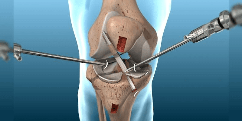
For meniscal tears: Arthroscopic treatment of the meniscal pathology — repair or partial meniscectomy — addresses the source of intra-articular inflammation. Baker's cysts associated with meniscal tears frequently resolve completely after arthroscopic meniscal surgery.
For RA: Systemic RA management (DMARDs, biologics) that achieves good disease control significantly reduces synovitis and with it the fluid accumulation driving the cyst.
Intra-Articular Steroid Injection
For OA-related Baker's cysts, an intra-articular corticosteroid injection into the knee joint — not the cyst — reduces synovitis, reduces fluid production, and typically results in the cyst reducing in size and often resolving over 4 to 8 weeks. This is more logical than aspirating the cyst directly, because it targets the source.
Aspiration of the Cyst: When It Is Appropriate
Aspiration is appropriate when:
- The cyst is very tense and causing significant symptoms (pressure, limited flexion)
- As a diagnostic procedure to confirm the fluid is benign
- Combined with an intra-articular steroid injection (treating the source at the same time)
Aspiration alone, without injection and without investigation of the underlying cause, should not be the standard management.
Surgical Excision: Rarely Needed
Surgical excision of a Baker's cyst is rarely indicated and rarely durable if the underlying joint pathology is not simultaneously addressed. Exceptions include very large cysts causing significant compression symptoms, or cysts with internal complexity suggesting a different diagnosis. Even when surgical removal is performed, recurrence is common unless the intra-articular cause is managed.
Baker's Cyst Complications: Rupture
Occasionally a large Baker's cyst ruptures — the fluid escapes from the cyst into the surrounding soft tissues of the calf. This causes sudden onset calf pain, swelling extending down from the knee into the calf, and sometimes bruising. The clinical picture can closely resemble deep vein thrombosis.
The important distinction: A ruptured Baker's cyst does not cause DVT, but it can look identical on clinical assessment. Ultrasound distinguishes the two — showing fluid tracking from the ruptured cyst in the case of rupture, versus venous thrombosis in DVT. If there is any doubt, both possibilities should be investigated.
A ruptured cyst is treated conservatively — rest, elevation, compression, and management of the underlying joint — and typically resolves over 2 to 4 weeks. Surgery is not needed for a ruptured Baker's cyst.
Frequently Asked Questions
Is a Baker's cyst dangerous? No — Baker's cysts are benign and do not become cancerous. The concern is the underlying joint problem causing it, not the cyst itself. The rare exception is if the clinical picture suggests something other than a simple Baker's cyst — which ultrasound investigation clarifies.
Can a Baker's cyst resolve on its own? Sometimes, particularly if the underlying cause temporarily resolves (an acute inflammatory flare settles, a mild meniscal tear becomes quiescent). In patients with chronic OA or untreated meniscal pathology, the cyst tends to persist until the underlying problem is managed.
My Baker's cyst keeps coming back after draining. What should I do? Request investigation of the underlying cause. An MRI of the knee to assess the articular cartilage, menisci, and synovium will identify why the knee is producing excess fluid. Treating the underlying pathology — not just repeatedly draining the cyst — is what breaks the recurrence cycle.
Dr. Ankur Singh | Knee Specialist Noida | Baker's Cyst Treatment India | Popliteal Cyst Noida | Sarvodaya Healthcare Noida | Renew Orthopedic Clinic Sector 47 Noida
Medical Disclaimer
The information provided on this website is for educational purposes only and should not be considered as medical advice. Please consult Dr. Ankur Singh or a qualified healthcare professional for personalized medical guidance.
Share this blog:
 Copy





/healthcare-concept-clinic-compressed.jpg)