What Happens During Knee Replacement Surgery: A Step-by-Step Guide

An operation is taking place in an operating room with surgeons working on a person's knee.
Most patients heading into knee replacement surgery have one significant anxiety that has nothing to do with pain or recovery — it is the simple not knowing. The operating theatre is a room most people have never seen. The procedure is a sequence of events most people cannot visualise. The unfamiliar is consistently more frightening than the understood.
This guide eliminates that unfamiliarity entirely. Everything that happens from the moment you arrive at KDSG Superspeciality Hospital in Greater Noida to the moment you wake up in the recovery room is described here, in sequence, in plain language. No medical jargon without explanation. No detail skipped.
By the time you finish reading, the operating theatre will be a familiar environment rather than an unknown one.
The Morning of Surgery
You arrive at KDSG Hospital at the time instructed by Dr. Ankur Singh's team — typically 6 to 7 in the morning for a morning operation. You have been fasting since midnight (water until 2 hours before surgery is usually permitted).
At the admission desk, the ward team takes you to the pre-operative bay — a preparation area adjacent to the operating theatres. A blood pressure cuff, oxygen probe, and heart monitor are connected. A nurse confirms your name, the operation being performed, and which knee is being operated. You will be asked this question multiple times by different staff — this is a safety protocol, not carelessness.
An intravenous (IV) line is placed in the back of your hand or forearm. Through this line, a pre-operative antibiotic is given — this critical infection prevention step happens before any skin incision. The antibiotic needs to be in your bloodstream before the wound is opened.
The anaesthesiologist visits you here. They review your medical history, confirm the plan (almost always spinal anaesthesia for knee replacement), and answer any last questions.
The Anaesthetic
For knee replacement surgery at KDSG, spinal anaesthesia is the standard technique — and it deserves understanding because most patients are unfamiliar with it.
Spinal anaesthesia (also called a spinal block) means that the anaesthetic medication is injected into the fluid surrounding the spinal cord in the lower back. This produces complete numbness from the waist downward within 3 to 5 minutes — you will feel nothing from the waist down for the duration of the surgery.
You are awake — or more precisely, you are drowsy and comfortable because a separate sedative medication is given through the IV line. Most patients describe this as being in a very relaxed, dreamlike state where they are aware of being in the theatre but feel no pain, no pressure, and no anxiety. Some patients drift into a light sleep.
Why spinal rather than general anaesthesia? Spinal anaesthesia avoids the risks of general anaesthesia (breathing tube complications, nausea, respiratory issues in elderly patients) and provides better post-operative pain control. It is safer for most knee replacement patients, particularly those with diabetes or cardiac conditions.
What you will feel: The spinal injection itself feels like a brief stinging and pressure sensation at the injection site. Within minutes, the legs become warm, then heavy, then completely numb. You will not be able to move your legs — this is expected and temporary.
Inside the Operating Theatre
The Environment
The operating theatre at KDSG is a laminar flow room — the air is filtered and circulated in a precisely controlled way to remove airborne bacteria and reduce infection risk. It is cooler than you might expect, brightly lit with overhead lights focused on the operating field, and filled with the organised activity of 4 to 5 people working together.
The surgical team consists of Dr. Ankur Singh (operating surgeon), an assistant surgeon, a scrub nurse (who handles the sterile instruments), a circulating nurse, and the anaesthesiologist who remains with you throughout.
Positioning
You are positioned lying on your back on the operating table. The operated leg is placed in a specific position using a leg holder that keeps the knee accessible and stable. A tourniquet is applied to the upper thigh — when inflated, this temporarily reduces blood flow to the leg, significantly reducing bleeding during the procedure. You feel nothing because of the spinal block.
The operated leg is cleaned thoroughly with antiseptic solution. Sterile drapes are placed over the leg, leaving only the knee area exposed. At this point, you are separated from the surgical field by a screen — you cannot see the surgery itself, which is intentional and preferable for most patients.
Step 1: The Incision (3 to 5 minutes)
A vertical incision is made over the front of the knee, typically 8 to 12 centimetres long. Skin, fat, and the joint capsule are opened in layers. This is the entry point to the joint.
Step 2: Exposing the Joint (5 to 8 minutes)
The kneecap (patella) is gently moved to one side — not removed, just displaced temporarily — to expose the full joint surface underneath. Dr. Ankur Singh can now see directly the worn cartilage surfaces: the end of the femur (thigh bone) and the top of the tibia (shin bone). The extent of cartilage damage is confirmed visually.
Step 3: Preparing the Bone Surfaces (20 to 30 minutes)
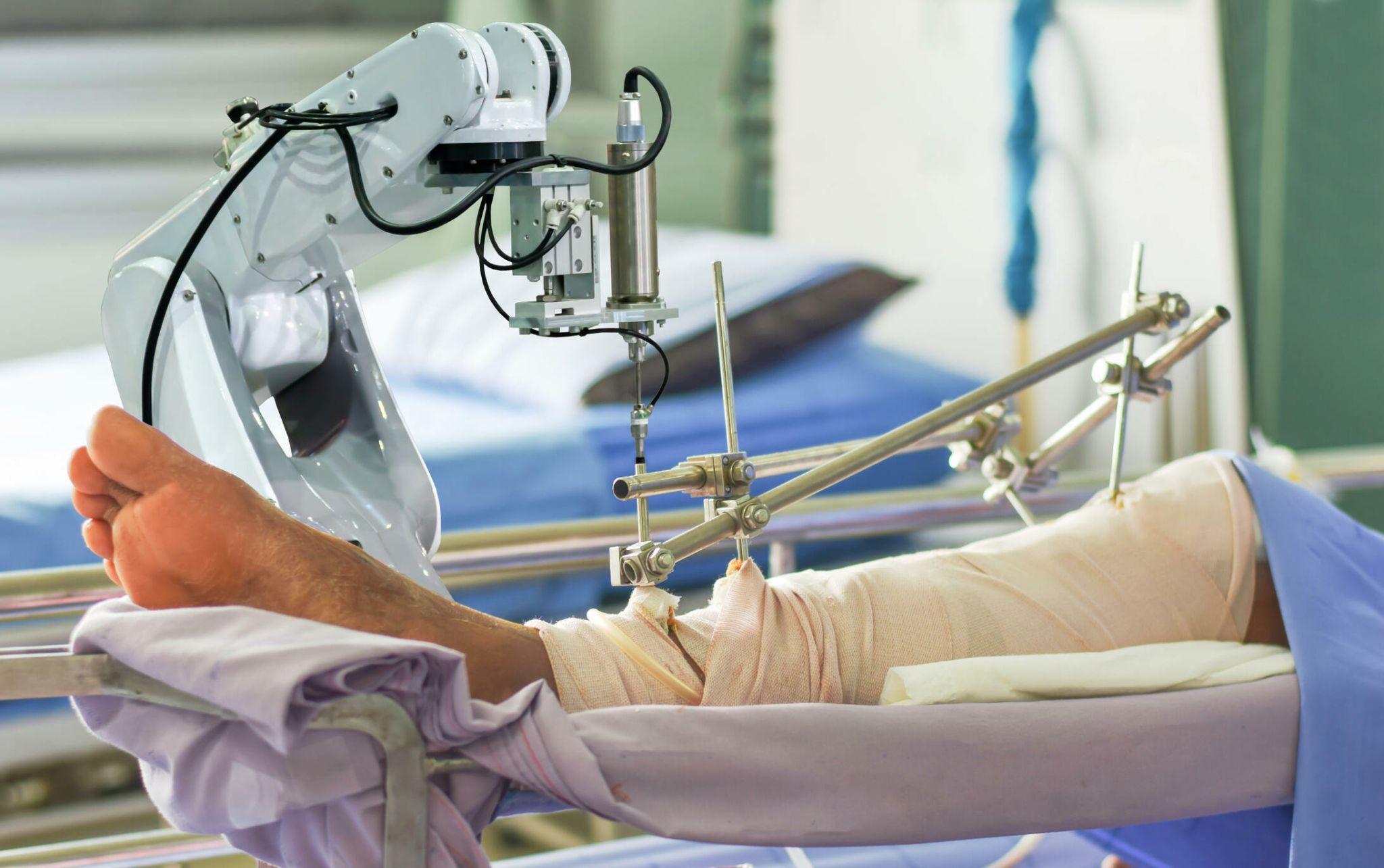
This is the most technically demanding phase of the operation. Using precision cutting guides — or, in a robotic procedure, robotic guidance that ensures each cut is within the pre-operatively planned zones — the arthritic bone surfaces are removed.
Femoral cuts: The end of the thigh bone is shaped to receive the femoral component — the metal cap that will cover the rounded end of the femur. Multiple careful cuts are made at precise angles and depths.
Tibial cuts: The top surface of the shin bone is cut flat and prepared to receive the tibial baseplate.
Patella preparation: In many knee replacements, the undersurface of the kneecap is also resurfaced with a polyethylene (plastic) button to improve kneecap movement over the new joint.
In a robotic-assisted procedure, each bone cut is verified against the pre-operative 3D plan in real time. The robotic system confirms that the bone has been cut to within 1 to 2 degrees and 1 to 2 millimetres of the planned target before proceeding.
Step 4: Trial Fitting and Alignment Check (10 minutes)
Before the final implant is placed, trial (temporary) components are fitted to the bone surfaces. The knee is moved through its full range of motion. Dr. Ankur Singh assesses alignment, stability, and the balance of the soft tissue tensions — ensuring the new joint is neither too tight nor too loose in flexion and extension.
This is a critical quality check. Any fine adjustments to the bone cuts or soft tissue are made at this point, before anything is permanently cemented.
In robotic surgery, the system also confirms the trial position against the plan — providing digital verification that the alignment target has been achieved.
Step 5: Implant Placement (10 to 15 minutes)
The trial components are removed. The final implants are placed.
The metal femoral component and the metal tibial baseplate are typically secured with bone cement — a fast-setting polymer that locks the metal to the bone within minutes. The polyethylene tibial insert (the plastic component that acts as the cartilage substitute) clicks into place on the tibial baseplate. If the kneecap is being resurfaced, the patella button is cemented onto the back of the kneecap.
The knee is again moved through its range of motion with the final implants in place to confirm optimal function.
Step 6: Wound Closure (15 minutes)
The joint capsule is closed with sutures, restoring the knee's natural envelope. The fat layer and skin are closed in separate layers. A dressing is applied. A drain tube may be placed to remove blood accumulating in the joint space over the next 24 hours — this is removed on the following day.
The tourniquet is released. Blood flow returns to the leg.
Recovery Room: 1 to 2 Hours
You are transferred to the recovery room immediately after the surgical team finishes. Recovery room nurses monitor:
- Blood pressure, heart rate, and oxygen levels
- Pain — medication is given through the IV as needed
- The wound dressing — checked for any excessive bleeding
- Your level of consciousness as the sedation fully wears off
The spinal anaesthesia begins to wear off over 2 to 4 hours. First you notice pins and needles. Then warmth. Then the ability to move the foot. Then the ability to flex and extend the knee.
Pain medication is administered proactively — before the spinal block completely wears off — so you do not experience a sharp transition from no pain to severe pain.
Once you are fully awake and observations are stable, you are taken back to the orthopaedic ward.
The First 24 Hours After Surgery
By the evening of surgery day or the morning after, the physiotherapist visits. The first exercises begin — ankle pumps, quadriceps sets — within hours of waking from surgery. The first standing and walking attempt happens on Day 1.
This is where the instinct of every patient fights the clinical evidence. Rest feels right. Walking feels impossible. The clinical evidence says: move. The patients who begin moving Day 1 consistently achieve better outcomes.
Frequently Asked Questions
Will I feel anything during the surgery?
With spinal anaesthesia, you will feel no pain or pressure below the waist. The sedation through the IV line keeps you drowsy and comfortable. Most patients report that the experience is far less distressing than they anticipated.
Can I watch the surgery?
A screen separates you from the surgical field and most patients prefer not to watch. In specific cases — and with the team's agreement — it is possible to observe. Most patients find the drowsiness from sedation makes this academic.
What if the spinal doesn't work completely?
This is very rare with experienced anaesthesiologists. If the spinal block is incomplete, additional anaesthetic can be given through the same injection point, or the anaesthetic plan can be converted to general anaesthesia. This decision is made entirely by the anaesthesiology team based on patient safety.
How do I know which leg will be operated?
Multiple verification checks occur before surgery — verbal confirmation, written marking on the skin, surgical site verification by the whole team in the OT. This multi-step process is the reason why wrong-site surgery, while receiving media attention, is extraordinarily rare in well-run hospitals.
Dr. Ankur Singh | Best Knee Replacement Surgeon in Noida | Knee Replacement Procedure Explained | KDSG Superspeciality Hospital Greater Noida | Renew Orthopedic Clinic Sector 47 Noida
Medical Disclaimer
The information provided on this website is for educational purposes only and should not be considered as medical advice. Please consult Dr. Ankur Singh or a qualified healthcare professional for personalized medical guidance.
Share this blog:
 Copy
Copy