Avascular Necrosis (AVN) of the Hip: The Complete Guide for Indian Patients

AVN typically strikes adults in their 30s and 40s, often after long courses of steroids.
Avascular necrosis (AVN) of the hip — also called osteonecrosis of the femoral head — is a condition in which the blood supply to the ball of the hip joint is disrupted, causing the bone to die. If caught early, the hip can be saved. If left until the femoral head collapses, hip replacement becomes unavoidable.
India has disproportionately high rates of AVN compared to Western countries. While osteoarthritis of the hip is the most prevalent indication for total hip replacement in the USA, AVN is one of the leading indications in India and many Asian countries. The reasons for this are specific to India's medical landscape — and understanding them matters for every patient who has received a steroid injection, had COVID-19, drinks alcohol, or has been on long-term medications.
This guide covers what AVN is, why it is so common in India, how it is staged, and what the treatment options look like at each stage — including the hip-preserving surgeries that can save the joint if intervention comes early enough.
What Is Avascular Necrosis of the Hip
The femoral head — the ball that fits into the hip socket — has a precarious blood supply. Unlike most bones, which have multiple routes of blood delivery, the femoral head relies primarily on a single set of vessels (the medial femoral circumflex artery branches) that travel along the femoral neck. When these vessels are damaged or blocked, a segment of the femoral head loses its blood supply.
Without blood, the bone cells (osteocytes) in the affected segment die. Dead bone loses its mechanical strength. Under the repetitive stress of body weight — every step, every stair — the weakened femoral head progressively collapses, destroying the smooth ball-and-socket architecture of the hip joint.
The tragedy of AVN is its typical patient: a person in their 30s or 40s, previously healthy, often with no hip pain at all in the early stages — who presents with suddenly severe hip pain when the femoral head has already begun to collapse.
Why AVN Is So Common in India: The Specific Risk Factors
Corticosteroid Use — The Leading Cause
Corticosteroid medications (prednisolone, methylprednisolone, dexamethasone) are among the most widely prescribed medications in India — for asthma, allergic conditions, skin diseases, nephrotic syndrome, joint conditions, and across multiple specialties. India's corticosteroid prescribing rates are substantially higher than in many Western countries, partly because access to newer biological medications is more limited, and partly because steroids are effective, inexpensive, and familiar.
Steroids cause AVN through two mechanisms: they increase blood lipid levels (fat emboli blocking the tiny vessels in the femoral head) and they cause direct toxic effects on osteocytes. The most common etiology of non-traumatic avascular necrosis in India is steroid intake, followed by idiopathic causes.
The cumulative dose matters — AVN risk rises significantly above approximately 2,000 mg of prednisolone equivalent, though it can occur at lower doses in susceptible individuals.
COVID-19 and Steroid-Related AVN — A Modern Epidemic
The COVID-19 pandemic generated a specific, large wave of AVN cases across India. Corticosteroids — particularly high-dose dexamethasone — were widely used to manage severe COVID-19 pneumonia. Post-COVID AVN of the hip is a delayed yet potentially preventable sequela associated with corticosteroid exposure and COVID-related vascular injury. The mean latency of 126 days from infection to AVN onset suggests a delayed presentation.
A Hyderabad retrospective study of 118 patients (212 hips) with post-COVID AVN found a mean age of 36.8 years with a majority being male (87.3%). The majority presented with bilateral AVN (79.7%), at Ficat and Arlet Grade 2 (50.9%) and Grade 3 (41%).
This means a large cohort of young Indian adults who received steroid treatment for COVID-19 in 2020 and 2021 developed bilateral hip AVN in their late 30s and early 40s — many of whom are now presenting with advanced disease or already requiring hip replacement.
Alcohol Use
Alcohol causes AVN through mechanisms similar to steroids — fat emboli and direct bone cell toxicity. Heavy alcohol use (more than 400 ml of spirits per week, or equivalent) significantly increases AVN risk. The Hyderabad study showing male predominance in COVID-related AVN also reflects the higher rates of alcohol use among Indian men.
Sickle Cell Disease
Sickle cell trait is present across central India and the Deccan plateau. Sickle-shaped red cells can block the microvasculature of the femoral head, causing AVN. Patients with known sickle cell disease should be specifically counselled about this risk.
Trauma and Hip Fracture
Femoral neck fractures — particularly displaced fractures — can disrupt the blood supply to the femoral head at the time of injury, causing post-traumatic AVN weeks to months later. This is the reason all significant hip fractures are assessed for AVN risk at follow-up.
Staging: Why It Determines Everything
AVN is staged by the Ficat and Arlet system (Stages 1 to 4), which corresponds to what is visible on X-ray and MRI. The stage at presentation determines what treatment options are available and what outcome is achievable.
Stage 1 — Normal X-ray, Abnormal MRI The femoral head looks normal on X-ray. MRI shows the area of dead bone as a well-defined lesion. No collapse has occurred. This is the stage where hip-preserving surgery is most likely to succeed. Pain may be mild or absent. Diagnosis is possible only with MRI — typically triggered by screening a patient with known risk factors (COVID steroid use, heavy alcohol use, transplant recipient).
Stage 2 — Abnormal X-ray, No Collapse X-ray shows density changes in the femoral head — sclerosis (whiteness), cystic areas. The femoral head shape is still intact. Hip-preserving surgery still possible with reasonable probability of success.
Stage 3 — Crescent Sign and Early Collapse The subchondral bone plate begins to separate from the underlying dead bone — producing the "crescent sign" on X-ray. The femoral head is beginning to collapse. Hip-preserving surgery has reduced success rates at this stage; some younger patients may still benefit.
Stage 4 — Femoral Head Collapse with Joint Space Loss The femoral head has deformed — the smooth ball is no longer round. The acetabulum (socket) develops secondary arthritis. At this stage, hip replacement is the standard treatment.
The critical clinical point: most patients present at Stage 3 or 4 because Stages 1 and 2 cause minimal pain. The pain becomes significant precisely when collapse begins — which is why opportunistic MRI screening in at-risk patients (COVID steroid recipients, heavy drinkers, patients on long-term oral steroids) is clinically important.
Treatment Options by Stage
Stage 1 and Early Stage 2: Joint-Preserving Surgery
Core Decompression
The most commonly performed hip-preserving procedure. One or more channels are drilled into the femoral head, reducing intraosseous pressure and theoretically allowing revascularisation — new blood vessel ingrowth into the dead segment. Core decompression is most effective for small lesions in Stage 1, where data shows approximately 60 to 70 percent survival of the native hip at 5 years.
Core decompression is often combined with bone grafting — packing the decompressed channel with bone graft material (autologous bone, allograft, or a synthetic substitute) to provide structural support to the softened femoral head.
Vascularised Fibular Graft
A technically demanding procedure where a segment of the patient's own fibula (calf bone), with its blood vessel still attached, is placed into the femoral head — providing both structural support and a new blood supply. Reserved for younger patients with larger lesions where core decompression alone is unlikely to suffice.
Stage 3 and Stage 4: Hip Replacement
Once significant collapse has occurred, the femoral head cannot be restored to its normal spherical shape. Total hip replacement — replacing both the femoral head and the acetabular socket — is the definitive treatment.
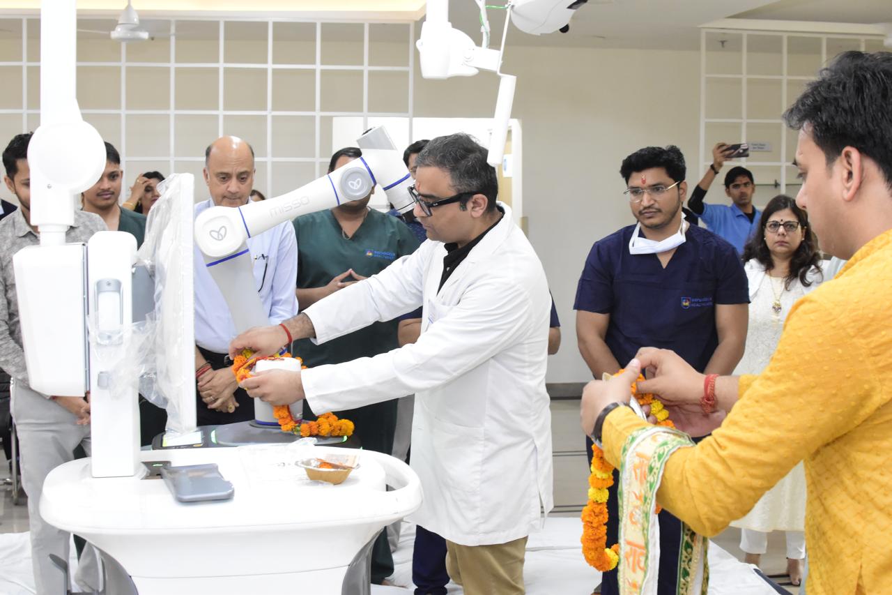
Hip replacement for AVN in India presents a specific challenge: the patients are typically much younger than the typical hip replacement patient (often mid-30s to mid-40s after COVID-related steroid AVN), meaning the implant needs to last 30 or more years. This makes implant selection and surgical precision particularly important — and is a strong argument for robotic-guided cup positioning in AVN-related hip replacement, which optimises the acetabular component orientation for long-term function.
Bilateral AVN: When both hips are affected — which is the case in the majority of post-COVID AVN patients — bilateral staged hip replacement may eventually be required. The first hip is typically done when symptoms are more severe; the second at a defined interval thereafter.
The Post-COVID AVN Patient: What Needs to Happen Now
If you received corticosteroids for COVID-19 in 2020 or 2021 and have not been screened for AVN, the question is not whether you need to worry — it is whether screening should have already happened.
Who should be screened with MRI: Any patient who received more than 2 weeks of corticosteroid treatment for COVID-19 and who is experiencing any hip or groin discomfort. The MRI should image both hips (bilateral screening).
Why early detection matters: The difference between Stage 1 diagnosis (where core decompression has a 60 to 70 percent chance of saving the hip) and Stage 3 to 4 diagnosis (where hip replacement is the only option) is typically a few months of symptom evolution. Early MRI is inexpensive relative to the difference in treatment complexity.
To book a consultation for AVN assessment or hip pain evaluation, call the number listed on this website. Consultations at Renew Orthopedic Clinic, Sector 47 Noida. Surgery at KDSG Superspeciality Hospital, Greater Noida.
Frequently Asked Questions
Can AVN heal on its own? AVN in Stage 1 can spontaneously stabilise in some cases — particularly with small lesion size. However, spontaneous progression to collapse is common enough that watchful waiting without treatment is generally not recommended for symptomatic Stage 1 or any Stage 2 disease. The risk of waiting and missing the window for hip-preserving surgery is real.
What if I received steroids for a joint injection rather than COVID? Joint injections (intra-articular steroids) carry substantially lower systemic exposure than oral or intravenous steroids. The AVN risk from isolated joint injections is very low. The risk is primarily from sustained systemic steroid courses — oral prednisolone taken for weeks to months.
How long does recovery take after hip replacement for AVN? Recovery from hip replacement for AVN is similar to that for osteoarthritis-related replacement — walking with a frame from Day 1, hospital discharge at 3 to 5 days, full daily activity by 3 to 4 months. The specific challenge for young AVN patients is long-term activity modification — protecting the new hip from high-impact activities to maximise implant lifespan.
Dr. Ankur Singh | Hip Replacement Specialist Noida Greater Noida | AVN Hip Treatment India | Post-COVID Avascular Necrosis | KDSG Superspeciality Hospital Greater Noida | Renew Orthopedic Clinic Sector 47 Noida
Medical Disclaimer
The information provided on this website is for educational purposes only and should not be considered as medical advice. Please consult Dr. Ankur Singh or a qualified healthcare professional for personalized medical guidance.
Share this blog:
 Copy
Copy