Hip Labral Tear: Causes, Symptoms, and Treatment Options for Patients in Noida

A man in a button-up shirt is standing with his back to the camera, one hand on his hip. A glowing illustration of the hip bone is overlaid, indicating pain or an issue in that area. The office environment in the background suggests a busy work life.
Hip labral tears are one of the more underrecognised causes of persistent groin and hip pain in younger adults — and one of the conditions that patients most frequently spend months or years trying to treat as something else before getting the right diagnosis.
The pain pattern is atypical enough that it often gets attributed to a muscle strain, a back problem, or just general hip tightness. Physiotherapy aimed at the wrong structure helps temporarily but not durably. The real problem — a tear in the acetabular labrum — remains unaddressed until someone orders the right investigation or refers to a specialist familiar with the condition.
This guide explains what the hip labrum is, how tears happen, what they feel like, how they are properly diagnosed, and what treatment involves for patients in Noida and Greater Noida.
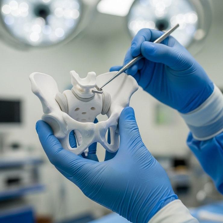
What Is the Hip Labrum?
The hip is a ball-and-socket joint — one of the most inherently stable joints in the body. The acetabulum (the socket) is deepened and surrounded by a ring of fibrocartilage called the acetabular labrum. This structure performs several important functions:
It deepens the socket, increasing the contact area between the femoral head (the ball) and the acetabulum. It creates a negative pressure "suction seal" that keeps the femoral head centred in the socket and helps distribute load evenly across the joint surface. It contains sensory nerve endings that provide proprioceptive feedback about hip position and movement.
When the labrum tears, this suction seal is compromised. The femoral head is no longer perfectly stabilised in the socket, and the load distribution across the joint changes — accelerating cartilage wear over time if left untreated.
How Does the Hip Labrum Tear?
Femoroacetabular Impingement (FAI)
This is the most common underlying cause of labral tears — accounting for the majority of cases seen in active adults in their 20s, 30s, and 40s. FAI occurs when there is abnormal contact between the femoral head-neck junction and the acetabular rim during hip movement, most commonly during flexion and internal rotation.
Two bony patterns drive FAI: CAM impingement (a bump at the head-neck junction of the femur), pincer impingement (overcoverage of the femoral head by the acetabular rim), or a combination of both. As the hip moves, this abnormal bony contact repetitively impacts the labrum at the site of impingement, causing cumulative damage that eventually results in a tear.
FAI-related labral tears are particularly common in athletes involved in sports requiring hip flexion and rotation — football, cricket (particularly batsmen and bowlers), martial arts, running, cycling, and gymnastics.
Traumatic Tears
A sudden, forceful hip movement — a fall, a twisting injury, or a hip dislocation — can tear the labrum acutely. Traumatic labral tears tend to produce immediate, sharp pain and may present alongside other hip injuries.
Hip Dysplasia
Hip dysplasia — where the acetabulum is shallower than normal, providing insufficient coverage of the femoral head — places the labrum under increased load because it is compensating for inadequate bony stability. Over time, this chronic overloading tears the labrum, often at multiple locations.
Degenerative Changes
In older adults, labral tears can occur as part of the broader degenerative process affecting the hip joint, often alongside early osteoarthritis.
Symptoms of a Hip Labral Tear
The symptom profile of hip labral tears is distinctive enough that an experienced clinician often suspects the diagnosis before imaging confirms it.
Groin pain. Deep groin pain — located at the front of the hip, felt deep in the crease where the thigh meets the pelvis — is the hallmark symptom. This pain is often described as a dull, deep ache that is present during activity and sometimes at rest.
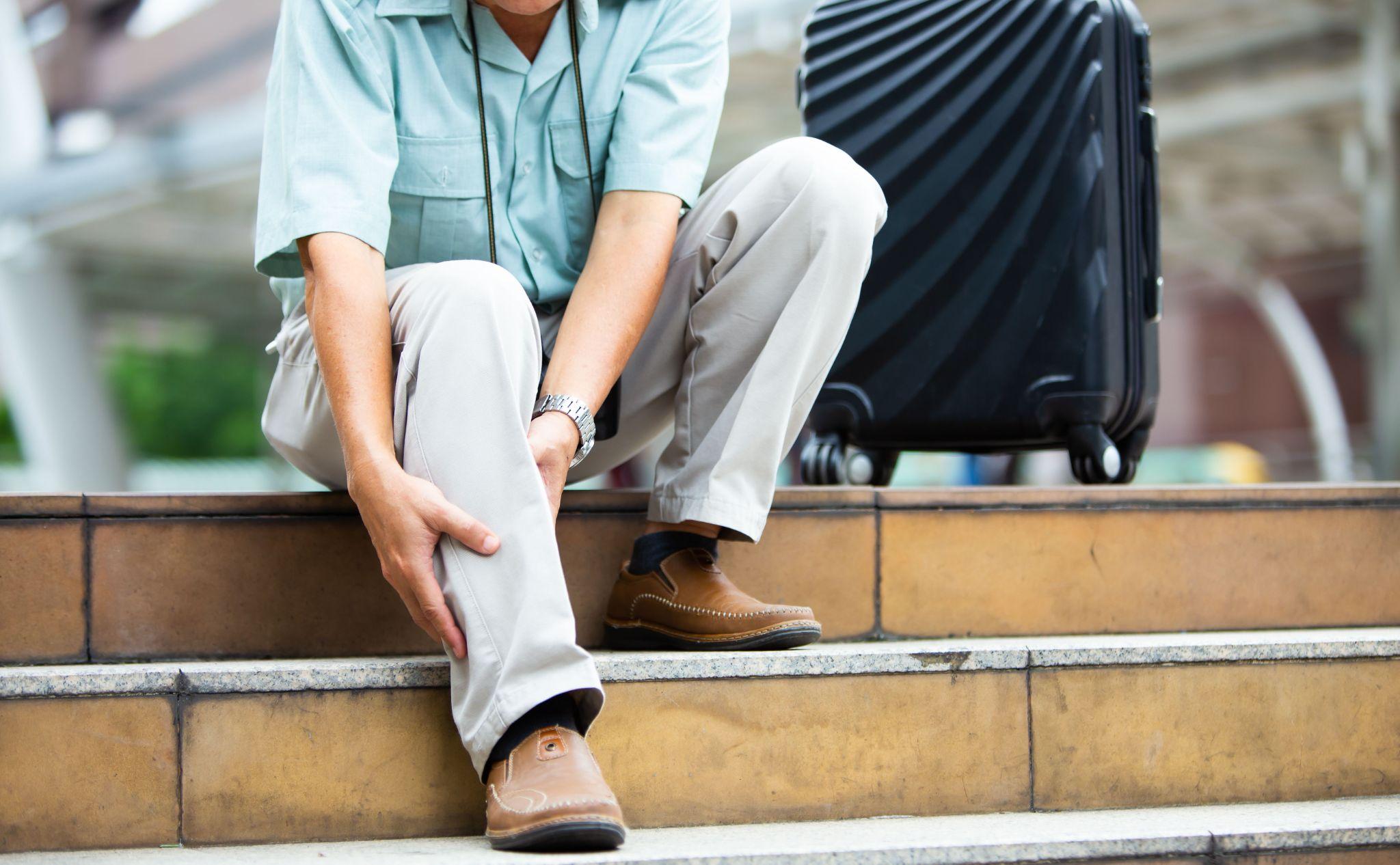
Pain with sitting. Many patients with labral tears find that prolonged sitting in a car, at a desk, or in a plane is particularly aggravating. The hip flexion position of sitting compresses the torn area of the labrum between the femoral head and the acetabular rim.
Clicking, catching, or locking sensation. Many patients describe a mechanical sensation inside the hip — a click, a catch, or a sense of the joint momentarily jamming during certain movements. This reflects the mechanical disruption of the labral seal.
Pain at the end of the range of motion. Reaching the limits of hip flexion (bending the hip fully) or internal rotation often reproduces a sharp pain or impingement sensation. This is a clinical sign that the surgeon specifically tests during examination.
Anterior hip or buttock pain. Pain may be felt at the outer hip or in the buttock, in addition to the groin. Patients sometimes describe using a "C-sign" — cupping their hand around the front of the hip to show where the pain originates.
Stiffness and reduced range of motion. The hip may feel stiff or restricted in certain directions, particularly internal rotation.
How Is a Hip Labral Tear Diagnosed?
Clinical Examination
An experienced hip specialist can often make a provisional diagnosis from the history and physical examination alone. Specific tests — the FADIR test (flexion, adduction, internal rotation) and the FABER test (flexion, abduction, external rotation) — reproduce the impingement and labral pain when positive. The combination of appropriate history and positive impingement signs points strongly to FAI with labral involvement.
X-Ray
Plain X-rays identify the bony morphology of the hip — the shape of the femoral head, the degree of acetabular coverage, and any cam or pincer deformity. They do not show soft tissue structures like the labrum itself.
MRI Arthrogram
The gold-standard imaging investigation for hip labral tears. A standard MRI has limited sensitivity for labral pathology. An MRI arthrogram — where a small amount of contrast dye is injected into the hip joint under imaging guidance before the MRI — dramatically improves the visibility of labral tears, cartilage damage, and other intra-articular structures. This is the investigation of choice when a labral tear is suspected clinically.
Treatment Options
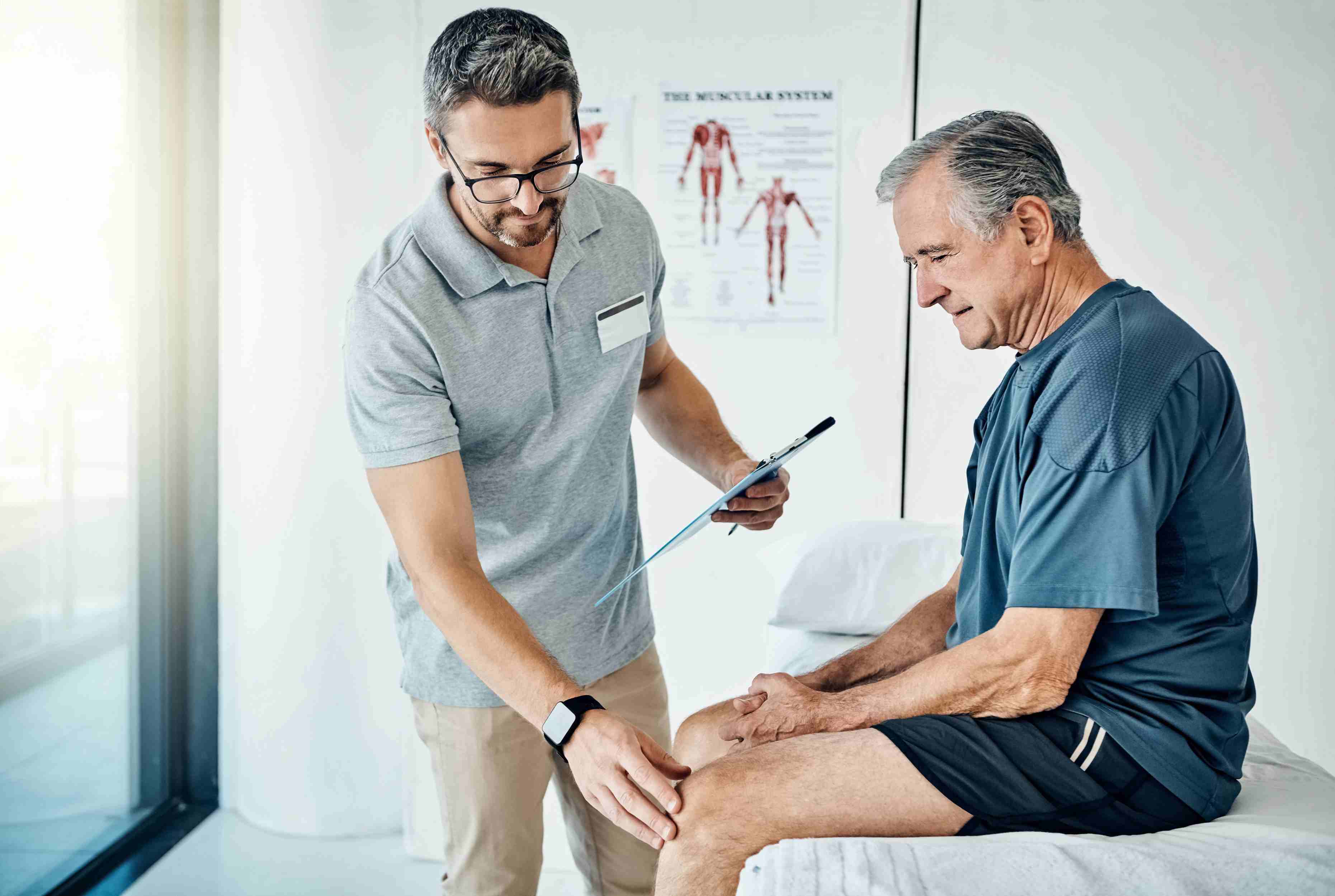
Conservative Management
Not every labral tear requires surgery. For patients with mild symptoms, small tears, or tears associated primarily with degenerative change, physiotherapy is the appropriate first-line treatment.
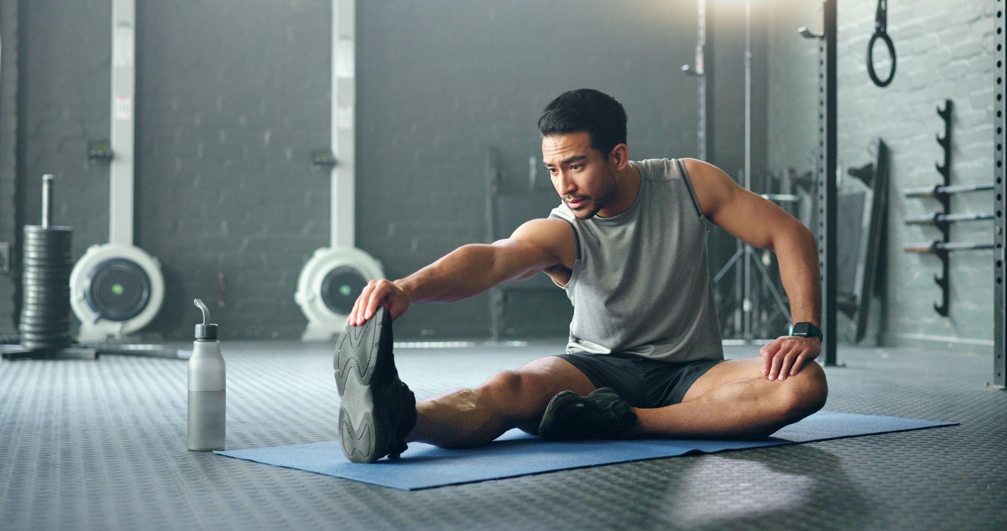
Physiotherapy for FAI and labral tears focuses on:
- Hip strengthening — particularly the gluteal muscles and hip external rotators — to reduce the dynamic impingement that aggravates the labrum
- Activity modification to avoid positions and movements that reproduce symptoms
- Core stability training to optimise pelvic control
- Movement re-education to avoid provocative patterns during sport and daily activity
A trial of three to four months of structured physiotherapy, combined with activity modification, is appropriate for most patients before surgical options are considered.
Corticosteroid injection into the hip joint, guided by ultrasound or fluoroscopy, can provide diagnostic confirmation and temporary symptom relief — particularly useful for patients whose symptoms are interfering significantly with daily life while they await surgery or pursue physiotherapy.
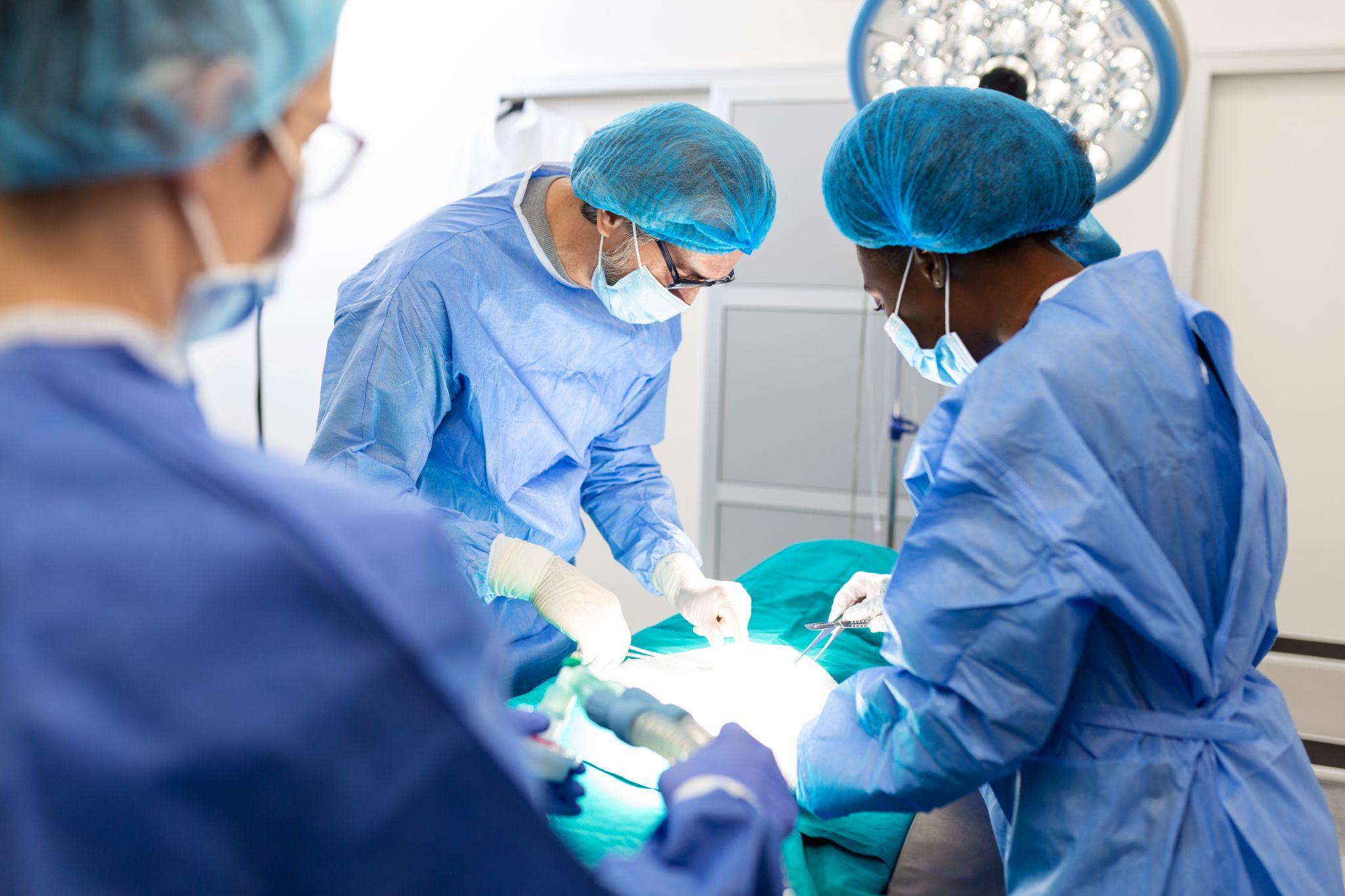
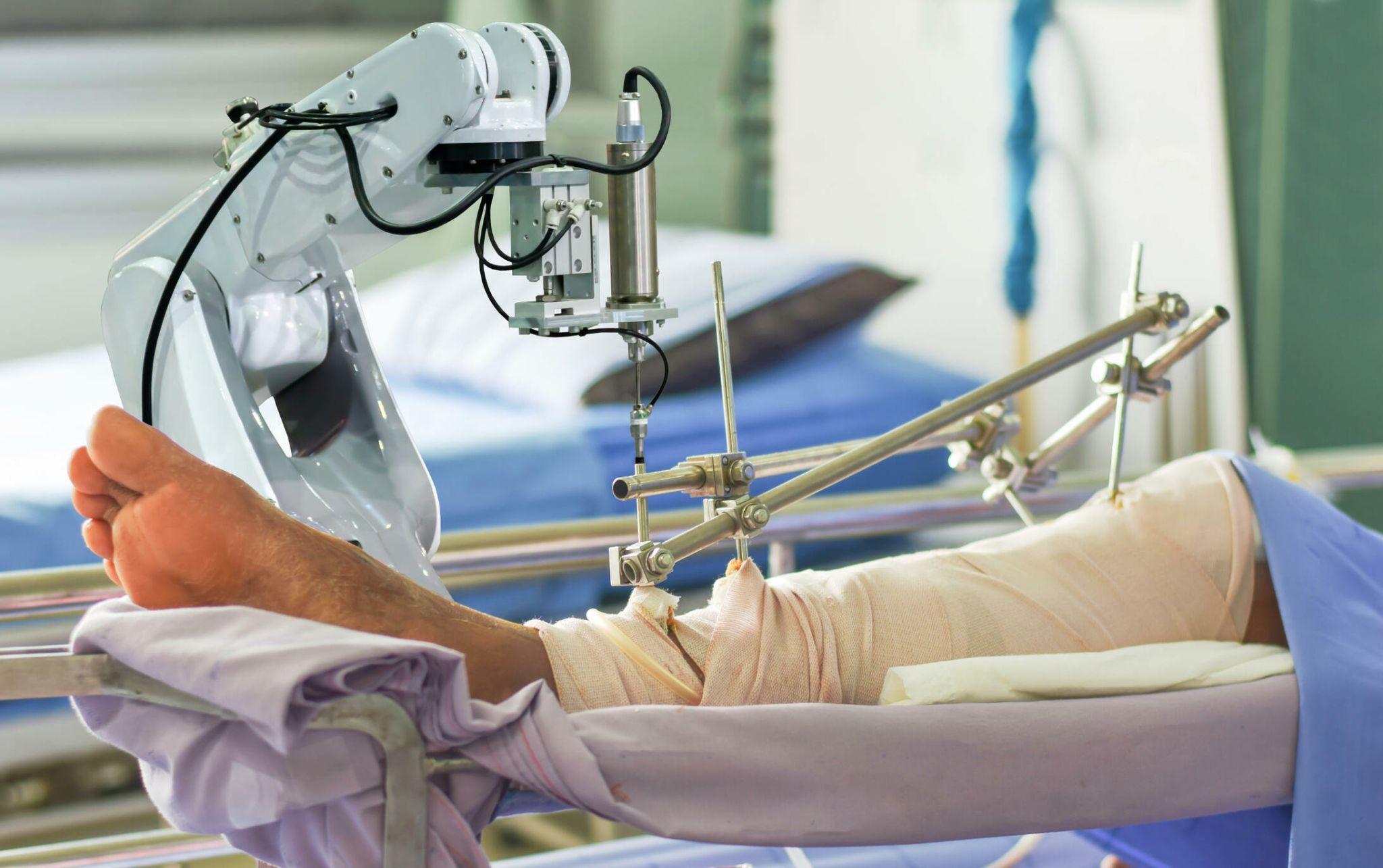
Hip Arthroscopy
When conservative management fails — or when the tear is large, the FAI is significant, or the patient is an athlete with specific return-to-sport demands — hip arthroscopy is the appropriate surgical intervention.
During hip arthroscopy, the surgeon makes two or three small incisions around the hip and inserts an arthroscope (camera) and specialised instruments. The procedure typically involves:
Labral repair: The torn labrum is reattached to the acetabular rim using small suture anchors. This is the preferred approach whenever the labrum is repairable, because preserving the labrum maintains the hip's suction seal and reduces the risk of progressive arthritis.
Cam/Pincer reshaping: If FAI is the underlying cause, the bony abnormality (the cam bump or acetabular rim overcoverage) is resected arthroscopically. Failure to address the underlying impingement leads to re-tear of the repaired labrum.
Cartilage treatment: Any associated cartilage damage is addressed at the same time — debridement, microfracture, or other cartilage techniques as appropriate.
Studies show that up to 89 percent of patients report clinical improvement after arthroscopic hip intervention when surgery is performed before significant arthritis has developed.
Recovery After Hip Arthroscopy
- Weeks 1–4: Partial weight-bearing with crutches. Gentle range-of-motion exercises.
- Weeks 4–8: Progressive weight-bearing. Physiotherapy intensifies.
- Months 2–4: Strengthening. Walking without a limp.
- Months 4–6: Return to non-contact sport.
- Months 6–12: Return to competitive sport for athletes.
Hip Labral Tear Care at Dr. Ankur Singh's Practice in Noida
Dr. Ankur Singh assesses and manages hip labral tears as part of his hip arthroscopy and joint preservation practice at KDSG Superspeciality Hospital in Greater Noida. Patients from across Noida, Greater Noida, and the Delhi-NCR region have access to specialist hip diagnosis and arthroscopic care without travelling to central Delhi.
If you have persistent groin or deep hip pain — especially with a clicking or catching sensation — and have not been evaluated for a labral tear, a specialist assessment is the right next step.
To book a consultation for hip pain evaluation in Noida or Greater Noida, call the number listed on this website.
Frequently Asked Questions
Can a hip labral tear heal on its own?
Labral tears generally do not heal spontaneously because the labrum has a limited blood supply. Symptoms may settle with rest and physiotherapy, but the structural tear remains. Whether this matters long-term depends on the tear's size, location, and the presence or absence of underlying FAI.
How long is the recovery after hip arthroscopy?
Most patients return to daily activities within 4 to 6 weeks, recreational activity by 4 to 6 months, and competitive sport by 9 to 12 months with proper rehabilitation.
Is hip arthroscopy available in Greater Noida?
Yes. Dr. Ankur Singh performs hip arthroscopy at KDSG Superspeciality Hospital in Greater Noida.
Does a labral tear lead to hip replacement?
If left untreated for many years, a labral tear with underlying FAI accelerates cartilage damage and can lead to early hip osteoarthritis. Timely surgical treatment prevents this progression in most patients.
Dr. Ankur Singh | Best Orthopedic Surgeon in Noida | Hip Labral Tear Treatment | Hip Arthroscopy Noida | KDSG Superspeciality Hospital, Greater Noida
Medical Disclaimer
The information provided on this website is for educational purposes only and should not be considered as medical advice. Please consult Dr. Ankur Singh or a qualified healthcare professional for personalized medical guidance.